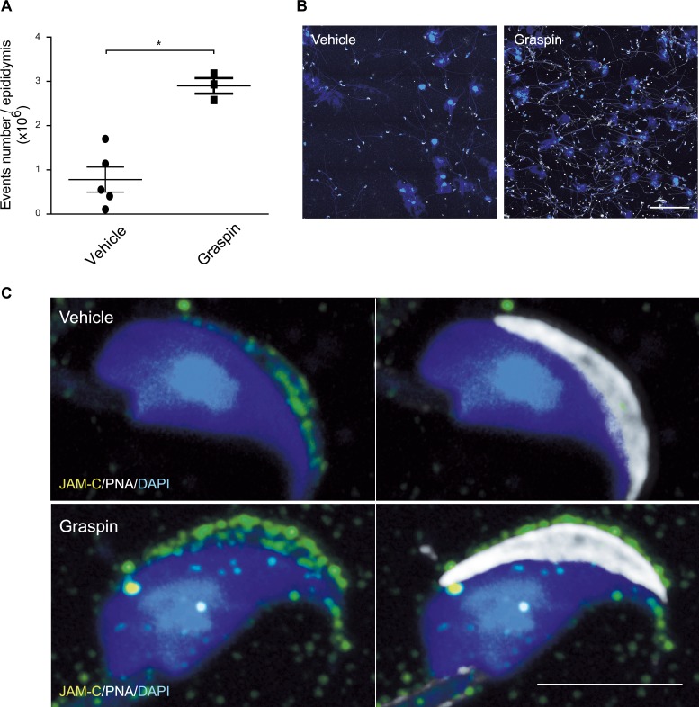
Fig 8. Graspin treatment induces germ cell release in epididymis.
(A) Quantification of epididymis content from vehicle and Graspin treated mice using flow-cytometry. Student’s unpaired t-test; *: P<0.05. (B) Cytospin of material recovered from epididymis of vehicle and Graspin treated mice and stained for DAPI and PNA. Note the increased abundance and heterogeneity of material recovered from Graspin treated animals. Scale bar, 100μm (C) Representative high magnification confocal images of spermatozoa recovered from epididymis of vehicle or Graspin treated mice and stained for DAPI (blue), JAM-C (green) and PNA (grey). Note that the co-localization of JAM-C with PNA is lost in samples from Graspin treated mice. Scale bar, 5μm

