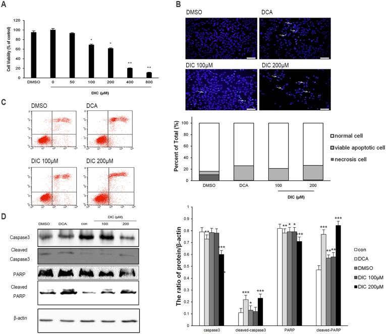
Fig 4. DIC inhibits cell viability and induces apoptosis in SKOV3 cells.
(A) SKOV3 cells were treated with increasing concentrations of DIC for 24 h, and cell viability was measured by the MTT assay. (B) After the indicated treatments for 24 h, SKOV3 cells were stained with Hoechst 33342 and imaged by confocal microscopy (scale bar, 50 μm; arrows, Hoechst-positive apoptotic cells). (C) Cells were treated as indicated for 24 h, stained with annexin V and PI, and analyzed by flow cytometry. Representative flow cytometry images are shown on the left, and the quantification of annexin V+PI+ apoptotic cells is shown on the right. (D) Upon treatment, the protein levels of caspase-3, cleaved caspase-3, PARP, and cleaved PARP in SKOV3 cells were examined by western blot, with representative western blot images presented on the left and the quantitation of protein expression relative to β-actin (internal control) presented on the right. Data are presented as the mean ± SE from three independent experiments, *P < 0.05, **P < 0.01, ***P < 0.001, when compared to nontreated control cells.

