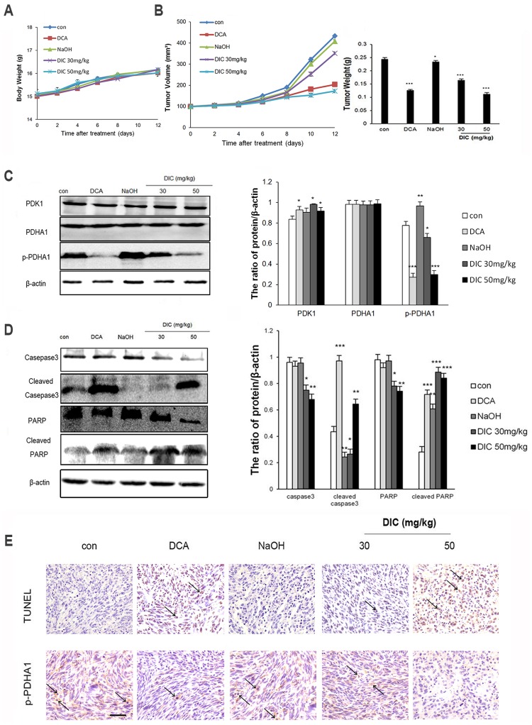
Fig 6. DIC suppresses tumor growth in vivo.
SKOV3 xenografts were established in nude mice, and the mice were treated as indicated. (A) The body weights of mice from all groups were monitored during the 12-day treatment period. (B) The tumor volume and tumor weight were measured at the indicated time points during treatment and at the time of sacrifice, respectively. (C) The protein levels of PDK1, PDHA1, and p-PDHA1 were examined by western immunoblotting, with β-actin examined as the internal control. Representative western blot images are shown on the left, and the quantification of protein levels relative to β-actin are shown on the right. (D) The protein levels of caspase-3, cleaved caspase-3, PARP, and cleaved-PARP in the SKOV3 xenografts were examined by western immunoblotting. Representative western blot images are shown on the left, and the quantification of protein levels relative to β-actin are shown on the right. Data are presented as the mean ± SE for all mice in each group. *P < 0.05, **P < 0.01, ***P < 0.001, when compared to the control group. (E) Detection of apoptosis and p-PDHA1 by the TUNEL assay and immunohistochemistry, respectively, in xenograft samples from the indicated groups (scale bar, 100 μm; arrows, apoptotic cells).

