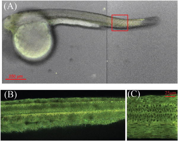
Fig. 6.

(A) Confocal image of zebrafish (48 hpf) at 10×magnification, after staining with flavonoid dye 2b. The images were taken from the head and tail sections. (B) and (C) Fluorescence confocal images of zebrafish from the tail section (approximate position is shown by a red square), at 20× and 100× magnification, respectively.
