
Figure 6. ACSL5, DGAT2, and CerS Interact on Lipid Droplet/ER interface.
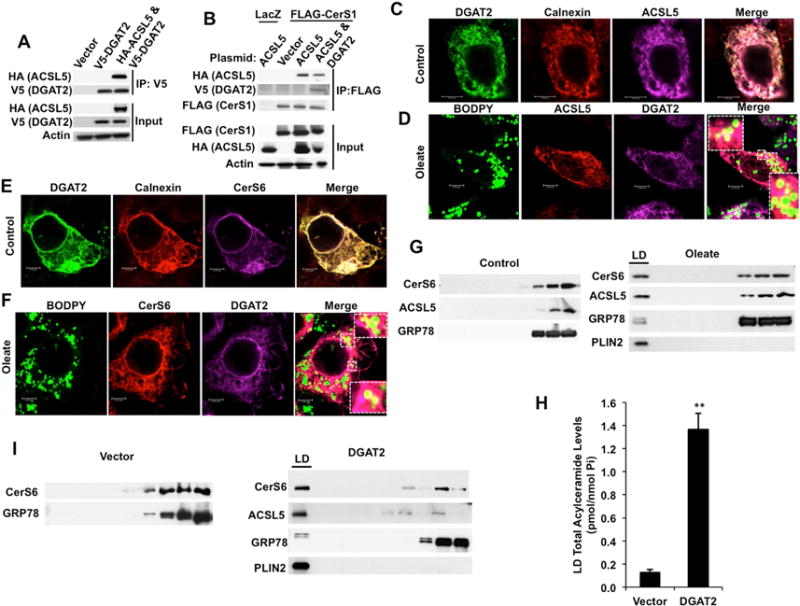
(A) Interaction of HA-tagged ACSL5 with V5-tagged DGAT2 was identified by anti-V5 IP and Western blotting with anti-HA and anti-V5 antibodies.
(B) Cells expressing FLAG-CerS1 were transfected with control vector, HA-ACSL5, or V5-DGAT2 in combination with HA-ACSL5 and formation of ACSL5-DGAT2-CerS1 complex was determined by anti-FLAG IP and Western blotting with anti-HA, -V5 and -FLAG antibodies. Results are representative of three independent experiments.
(C and D) Co-localization of HA-ACSL5 with V5-DGAT2 on ER (Calnexin) and lipid droplets (BODIPY) was imaged using confocal microscopy in the absence (C) and presence of 0.5 mM oleate (D). Inset: Magnified images of indicated areas.
(E and F) Co-localization of FLAG-CerS6 with V5-DGAT2 on ER (Calnexin) and lipid droplets (BODIPY) was imaged in the absence (E) and presence of 0.5 mM oleate (F). Inset: Magnified images of indicated areas.
(G) Localization of endogenous CerS6 and ACSL5 in lipid droplets upon oleate treatment was detected using cellular fractionation followed by Western blotting. LD: lipid droplet fractions.
(H) Total acylceramide content of lipid droplets isolated from cells stably expressing vector control or DGAT2 was measured by LC/MS.
(I) Localization of CerS6 in lipid droplet fractions was determined by Western blotting in cells stably transfected with control vector (left panel) or DGAT2 (right panel). LD: Lipid droplet fractions. Results are representative of at least three independent experiments.