
Fig. 7. WNT/β-catenin signaling pathway in BLCC-treated VLUs and correlation with healing trajectory.
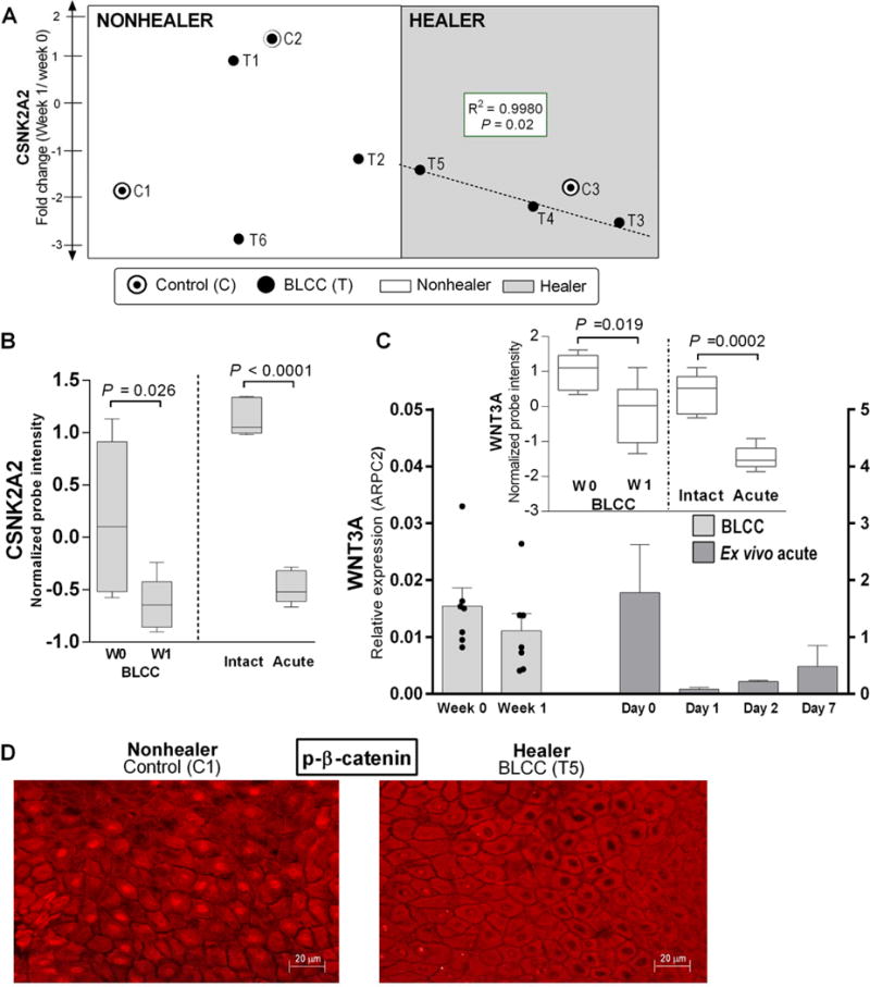
(A) CSNK2A2 expression changes and correlation with wound closure trajectory slope in 3 BLCC-responsive “Healers” (shaded area, dashed line) compared to 3 “Nonhealers” and 3 controls. See also fig. S5. (B) CSNK2A2 expression in healing acute wounds (n=6) and in chronic VLUs treated with BLCC (n=6), as represented by box-and-whisker plots of microarray probe intensity; paired t-test. (C) Relative WNT3 expression in chronic VLUs (n=7) before and after BLCC treatment and in acute wound healing (n=2 healthy donor skin, ex vivo assay). Bars represent mean and SEM of 3 technical replicates after normalization to ARPC2 internal control. Inset: WNT3 microarray probe expression in paired biopsies of chronic VLUs pre- vs. post-BLCC treatment (n=6) as well as acute wounds pre- vs. 3 days post-wounding (n=6); paired t-test P<0.05. (D) Immunofluorescence of phospho-β catenin cellular localization in keratinocytes at the wound edge of a “Nonhealer” VLU treated with standard of care (control), versus a BLCC-treated “Healer” VLU. Images are representative of 3 controls and 3 BLCC-treated VLU “Healers”. Scale bar 20μm.