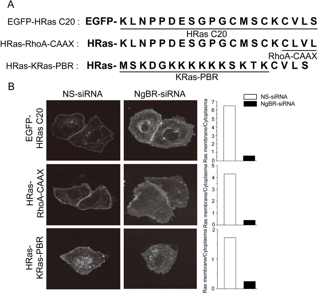
Figure 3. The CAAX motif of H-Ras is critical for binding NgBR.
(A) The diagram shows the H-Ras domain mutants. EGFP-HRas C20: EGFP fused to the C-terminal 20 residues of H-Ras; HRas-RhoA-CAAX: EGFP fused to full-length H-Ras that has its CAAX motif (CVLS) replaced with the RhoA CAAX motif (CLVL); HRas-KRas-PBR: EGFP fused to full-length H-Ras that has its aa 170 – 185 (klnppdesgpgcmsck) replaced with aa 170–185 of K-Ras constituting the polybasic region (mskdgkkkkkksktkc). (B) NgBR knockdown inhibited membrane localization of EGFP-HRasC20 and HRas-KRas-PBR. Images of EGFP-HRas membrane localization (left panel), and quantification of the results (right panel) are shown.

