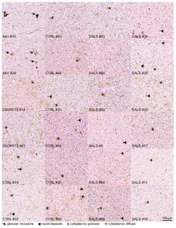
Fig. 3. No misfolded SOD1 is immunodetected in SALS patients using the 10E11 DSE2 misfolded SOD1 antibody.
Immunochemical analysis of misfolded SOD1 in two FALS patients with SOD1A4V mutation (#15 and #24), two with expansion repeats in C9ORF72 gene (#14 and #81), eight non-neurological controls (#19, 37, 83, 44, 23, 31, 20 and 65) and twelve SALS (#63, 60, 82, 6, 64, 68, 36, 35, 32, 17, 11, 16) using the 10E11 DSE2 antibody. Arrows indicate the presence of globular inclusions or intense positive aggregates of misfolded SOD1. Arrowheads point to immunoreactive round deposits found randomly, more frequently outside cells. The cross or asterisk signs indicate cytoplasmic diffuse or granular signal in neurons, respectively. Scale bar, 100 μm.

