
Figure 1.
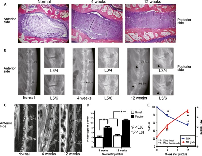
Altered histological, radiographic and magnetic resonance imaging (MRI) results of the intervertebral discs (IVDs) after needle puncture. (A) Typical sections stained with hematoxylin and eosin (H & E; 40 ×) are shown. (B) Radiograph showing a decrease in the L3–4 and L5–6 disc height accompanied by endplate sclerosis and the formation of vertebral osteophytes (black triangle). The L3–4 and L5–6 discs are enlarged in the right panel. (C) Decreased signal intensity of the punctured discs upon MRI imaging. (D) Histological changes in IVDs after puncture were evaluated using a grading score, and all data are presented as the mean ± standard deviation (SD). *P < 0.05, **P < 0.01, n = 6. (E) Decreased % disc height index (DHI) of the punctured discs and gradually increased MRI score using modified Thompson classification. All the data are presented as the mean ± SD. **P < 0.01, n = 6 discs from three rabbits.