
Figure 2.
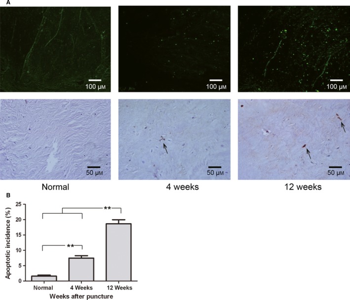
Apoptosis in the nucleus pulposus (NP) detected by TUNEL staining. (A) Apoptotic cells were visualized using fluorescence microscopy and light microscopy (200 ×, 400 ×). (B) Quantification of the apoptotic incidence between groups. Apoptosis varied significantly at each time point after puncture. **P < 0.01, values represent the mean ± SD of six discs from three rabbits.