
Figure 4.
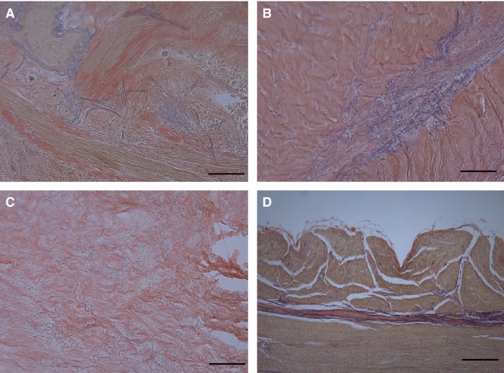
Weigert‐van Gieson stains of various fasciae to show arrangement of elastic fibres: lung fascia (A), visceral fascia of abdomen (B), liver fascia (C), oesophagus fascia (D). Scale bars: 150 μm (A–D). In visceral fascia of abdomen, elastic fibres are mainly found in loose connective tissue between fibrous sublayers.