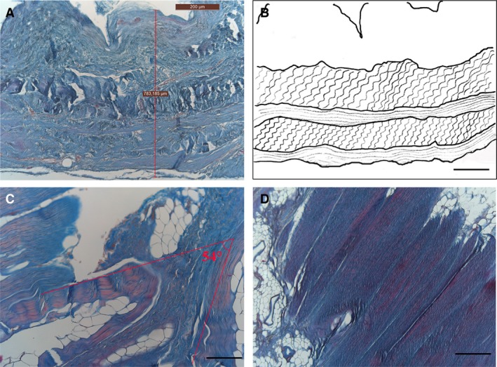
Figure 5.

Arrangement of collagen fibres in visceral fasciae, azan‐Mallory stain (A, C, D). (A) Full wall thickness of pericardial sac: note several fibrous sublayers. (B) Diagram of collagen arrangement. (C) Visceral fascia of thorax: note angle of approximately 54 ° between adjacent fibrous sublayers forming (C). Section of a sublayer of visceral fascia of abdomen, showing how collagen fibres are all arranged in the same parallel direction (D). Scale bars: 300 μm (A); 150 μm (B–D).
