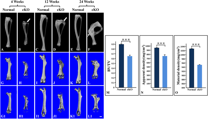
Figure 2.

X-ray radiography analyses. (A–F) Plain X-ray radiographs of mouse tibias; arrows in (B) and (D) were pointing at the growth plate zones; arrow in (F) was pointing at a healed fracture. (G–L) whole views of µCT scans for mouse femurs; (G1–L1) longitudinal section views of femurs; (M–O) quantitative analyses of data acquired from the high-resolution scans of the midshaft regions in the femurs from five mice (n = 5) at postnatal 12 weeks. Bar = 1.0 mm; ***P < 0.01. The long bones of cKO mice were shorter and had lower radiodensity compared to the age-matched normal mice (A–F). The growth plate zones (arrows in B and D) appeared wider in the cKO mice than in the normal mice. The tibia of 24-week-old cKO mice had a fracture (arrow in F), which appeared to have healed. The metaphysis regions of the femurs in the cKO mice were enlarged; the outer and inner bone surfaces of the long bones were more porous (G–L,G1–L1). The cortical bone of cKO mice had significant reductions in BV/TV ratio (M), Apparent Density (N) and Material Density (O).
