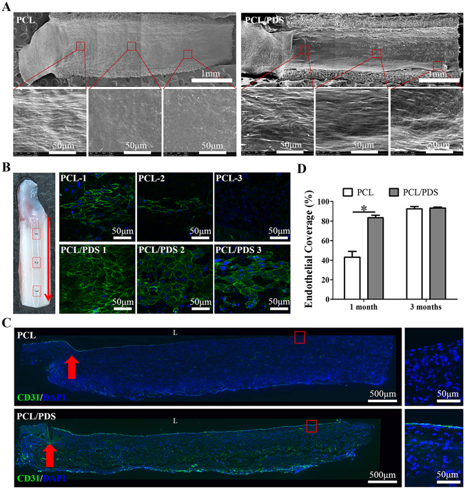
Figure 3.

Endothelialization formation of the explanted grafts at 1 month after implantation. (A) The lumen surface of explanted grafts was observed by SEM. (B) The endothelial coverage of grafts was observed by En face immunostaining using CD31 antibody (Red arrows: blood flow direction). (C) Endothelialization was analyzed by immunofluorescence staining of longitudinal sections of grafts using CD31 antibody (L: lumen; Red arrows: suture site). (D) The endothelial coverage at 1 and 3 months was calculated based on the longitudinal sections with CD31 staining. *p < 0.05.
