
Figure 1.
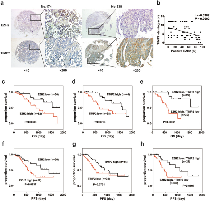
EZH2 inversely correlates with TIMP2 and indicates poor prognosis. (a) Representative images of immunohistochemistry analysis of EZH2 and TIMP2 in ovarian cancer tissues. EZH2 was expressed in the nucleus, while TIMP2 was expressed in the cytoplasm. Sample No. 174 has high EZH2 expression and low TIMP2 expression. Sample No. 338 has low EZH2 expression and high TIMP2 expression (scale bar, 50 μm). (b) Negative correlation between the percentage of EZH2-positive cells and the TIMP2 staining score in ovarian cancer tissues (Spearman’s correlation test). (c–e) Kaplan-Meier plots for overall survival in epithelial ovarian cancer patients. High EZH2 (c), low TIMP2 (d) or high EZH2 and simultaneous low TIMP2 (e) are associated with reduced overall survival. (f–h) Kaplan-Meier plots for progression-free survival in patients with epithelial ovarian cancer stratified by the expression status of EZH2 (f), TIMP2 (g), or both EZH2 and TIMP2 (h).