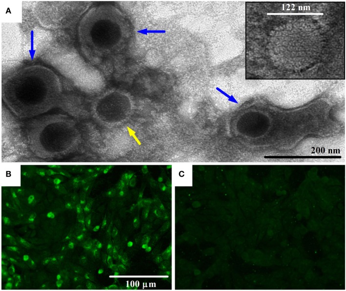
Figure 1.
Transmission electron microscopy (TEM) image of bovine gammaherpesvirus 4 (BoHV-4)-FMV isolate and detection of its antigens by indirect immunofluorescence assay (IFA). (A) TEM image of BoHV-4-FMV virus isolate grown on MDBK cells, showing herpesvirus-like virions. Negative-stain TEM allows the visualization of enveloped (blue arrows) and non-enveloped (yellow arrow) herpesvirus particles. A more detailed icosahedral capsid structure of the virus with an overall size of 122 nm in diameter, with its capsomere subunits, is illustrated in the inset. (B) Visualization of BoHV-4-FMV MDBK-infected cells by a specific IFA. (C) Mock MDBK-infected cells.

