
Figure 2.
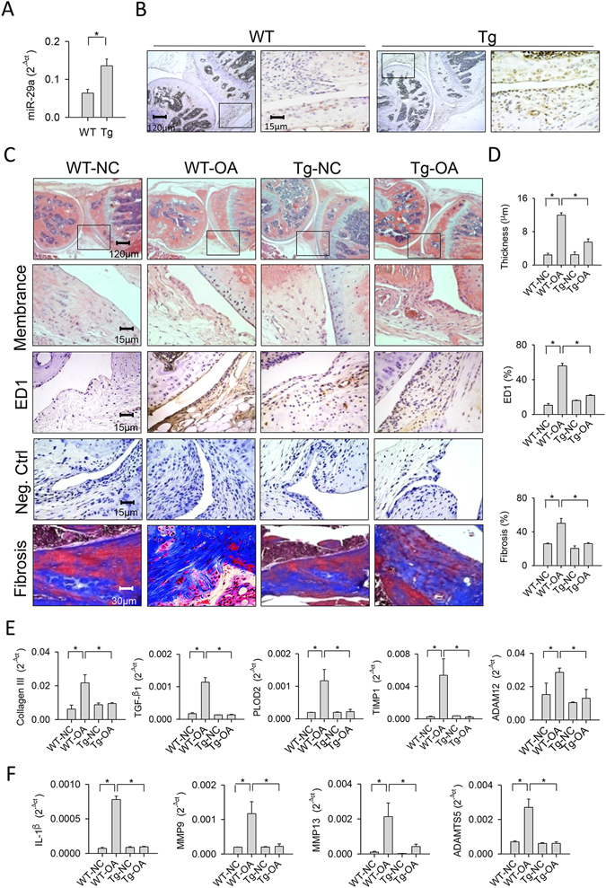
Analyses of synovial tissues in collagenase-affected joints in the miR-29aTg mice and wild-type mice. (A) Synovial tissues in the miR-29aTg mice showed high miR-29a expression probed by RT-qPCR and analyzed using a Wilcoxon test. (B) They also displayed strong miR-29a transcripts as evidenced by in situ hybridization. Panels in the low power field images indicate areas of interest for high power field images of synovial membrane. (C) Synovial tissues in the miR-29aTg mice exhibited slight hypertrophy, ED1-positive macrophage infiltration, and fibrotic matrix accumulation in collagenase-injured joints. (D) miR-29Tg mice had minor responses to collagenase aggravation of membrane thickness, macrophage number, and fibrosis tissue area. miR-29a overexpression reduced the collagenases enhancement of (E) collagen III, TGF-β1, PLOD2, TIMP1, ADAM12, (F) IL-1β, MMP9, MMP13, and ADAMTS5 expressions within injured joints. Each bar plot stands for mean ± standard error calculated from 8 animals in each group and analyzed by a parametric analysis of variance (ANOVA) and a Bonferroni post-hoc test. Asterisks (*) stands for significant difference between groups.