
Figure 3.
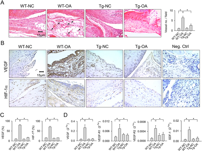
Analyses of angiogenic activities in synovial tissues. (A) Few vessels (arrows) were developed within collagenase-injured joints in the miR-29aTg mice. (B) Synovial tissues in the miR-29aTg mice displayed minor response to collagenase escalation of VEGF and HIF-1α immunostaining. (C) miR-29a overexpression reduced the number of synovial fibroblasts positive for VEGF and HIF-1α. (D) miR-29a reduced the collagenase elevation of VEGF, VEGF-R1, VEGF-R2, and SDF-1 expression in synovial tissues. Each bar plot stands for mean ± standard error calculated from 8 animals in each group and analyzed by a parametric analysis of variance (ANOVA) and a Bonferroni post-hoc test. Asterisks (*) stands for significant difference between groups.