
Figure 4.
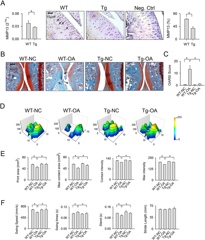
Cartilage morphology and gait characteristics of collagenase-affected joints in miR-29aTg mice and wild-type mice. (A) Articular chondrocytes in the miR-29aTg mice displayed weak MMP13 mRNA expression and immunostaining (arrows) in knee joints. (B) Injured knees in the miR-29aTg mice showed smooth articular cartilage morphology (arrows) and intense Safarin O staining in association with (C) lower OARSI scores than those of the wild-type mice. (D) The miR-29aTg mice showed regular footprint histograms in the collagenase-injected knees. The miR-29aTg mice exhibited minor response to the collagenase disturbance of (E) print area, maximum contact area, contact intensity, maximum intensity affected joints. (F) The collagenase disturbance of wing speed, swing time, and stand time were lessened in the miR-29aTg mice. Each bar plot stands for mean ± standard error calculated from 8 animals in each group and analyzed by a parametric analysis of variance (ANOVA) and a Bonferroni post-hoc test. Asterisks (*) stands for significant difference between groups.