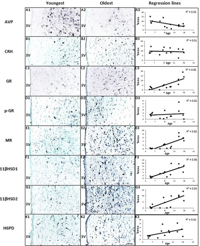
Figure 3. The effects of age on area% positive staining of HPAA proteins in hypothalamus of female baboons (P. hamadryas).
Columns A1-K1 and A2-K2 show the youngest and oldest photomicrographs. Column A3-K3 shows correlations between age and peptides of hypothalamus. Linear regression showed AVP expression was negatively correlated with age (R = −0.57, P < 0.05). GR, MR, 11βHSD1, and 11βHSD2 showed positive age-related regression in the PVN of hypothalamus (R values were as follows: 0.78, 0.79, 0.76, 0.73, P < 0.05, respectively). CRH and p-GR were not correlated with ages (P > 0.05). The expression of H6PD tended to correlate positively with age in the PVN of hypothalamus (R = 0.49, P = 0.08). Scale bar (100µm) applies to all micrographs.

