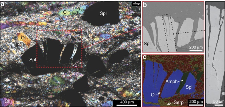
Figure 2. Mode-I cracks in spinel located near the centre of a mylonitic complex.
(a) Thin section (polarized light) of a mylonite layer containing cracked spinel (Spl; chromite) in between ultramylonite layers (a white star shows the field location in Fig. 1b). (b) Backscatter electron (BSE) image showing the mode-I cracks in spinel. See the methods section for analytical conditions. (c) Element map of the BSE image using energy-dispersive X-ray spectroscopy (EDX). The colour coding is based on the relative amounts of O, Mg, Si, Fe, Al, Ca and Cr. This map highlights olivine (Ol) and amphibole (Amph) as filling the mode-I cracks. Serp=serpentine.

