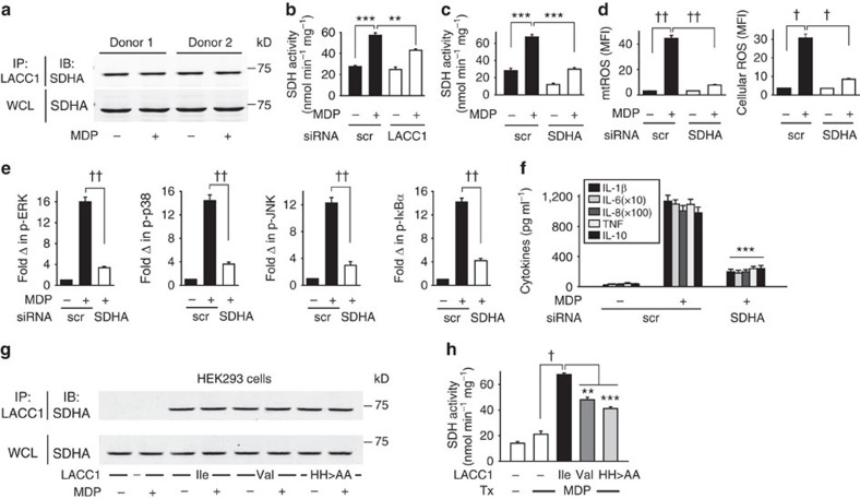
Figure 7. LACC1 associates with SDHA and contributes to NOD2-induced SDH activity.
(a) MDMs were left untreated or were treated with 100 μg ml−1 MDP for 6 h. Cell lysates were immunoprecipitated (IP) with anti-LACC1 antibody. Associated SDHA was assessed by western blot (IB). Representative western blot in two of four individuals. (b) MDMs (n=4) were transfected with scrambled or LACC1 siRNA, and then treated with 100 μg ml−1 MDP and assessed for SDH activity at 2 h. Similar results were observed in an additional n=8. In a subset of individuals we simultaneously confirmed reduced LACC1 expression with LACC1 siRNA by western blot (Supplementary Fig. 14A) and flow cytometry. (c–f) MDMs were transfected with scrambled or SDHA siRNA, and then treated with 100 μg ml−1 MDP and assessed for: (c) SDH activity at 2 h (n=4), (d) mtROS and cellular ROS at 6 h (n=4; similar results in an additional n=4), (e) fold phospho-protein induction normalized to untreated cells at 15 min (n=4) as assessed by flow cytometry, (f) cytokines at 24 h (n=4). (g,h) LACC1 Ile254, Val254 and His249,250Ala variants were transfected into HEK293 cells along with NOD2, and cells were then treated with 100 μg ml−1 MDP. (g) Cell lysates were immunoprecipitated (IP) with anti-LACC1 antibody at 6 h. Associated SDHA was assessed by western blot (IB). Shown is a representative western blot from one of three replicates. (h) SDH activity was assessed. Data represent three replicates and were repeated two independent times. Shown is mean+s.e.m. for (b–f,h). WCL, whole cell lysate; Tx, treatment; scr, scrambled. **P<0.01; ***P<0.001; †P<1 × 10⁻⁴; ††P<1 × 10⁻⁵; determined by 2-tailed t-test.

