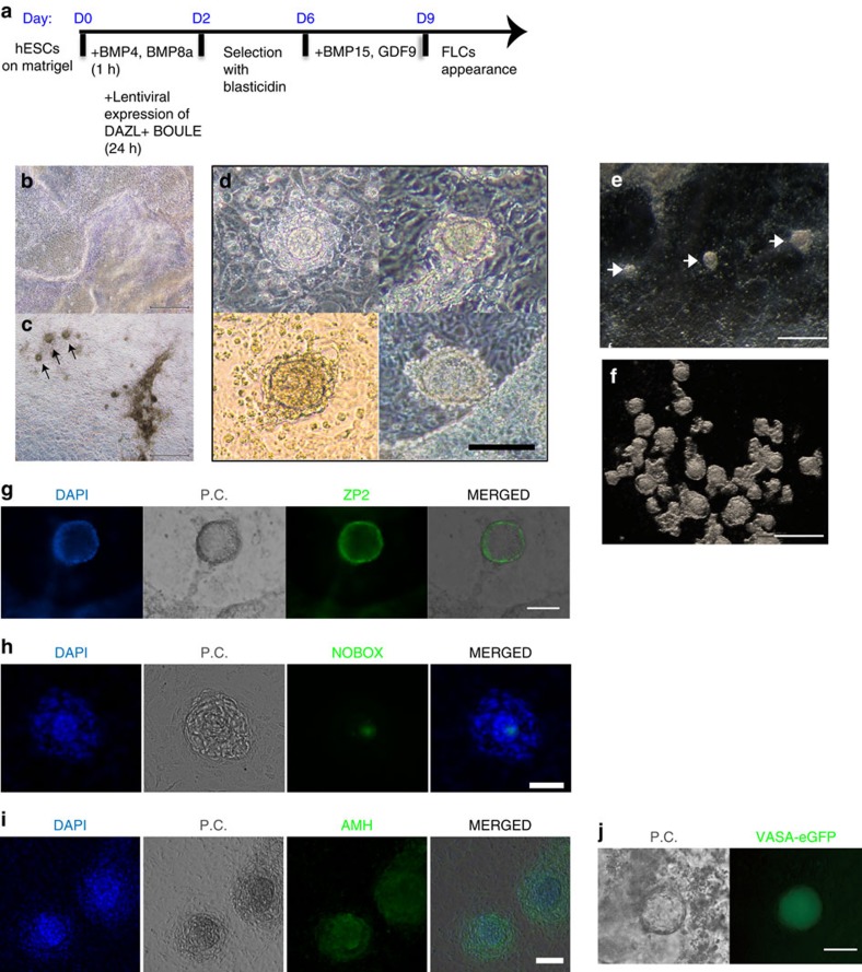
Figure 5. Appearance of ovarian FLCs induced in differentiated hESCs.
(a) Schematic timelines and simplified protocols for the differentiation from the undifferentiated hESC stage to the appearance of FLCs. (b) Phase-contrast image of a differentiated hESC culture without inducers at day 11. (c) Phase-contrast image of a differentiated hESC culture with the inducers described in a at day 11. (d) Magnified phase-contrast images of four different FLCs appearing at day 11 in the induced culture. (e) Stereo-microscope image of a differentiated hESC culture with inducers at day 11. (f) Stereo-microscope image of a group of FLCs collected from a differentiated hESC culture with inducers at day 11. Immunofluorescent staining of ovarian follicle markers ZP2 (g), NOBOX (h) and AMH (i) on the culture plate with FLCs. (j) VASA-eGFP reporter cells specifically expressed eGFP in cells that became FLCs. P.C.: phase contrast. Bar, 500 μm in b,c,e,f; 100 μm in d; 50 μm in g,h,i,j.

