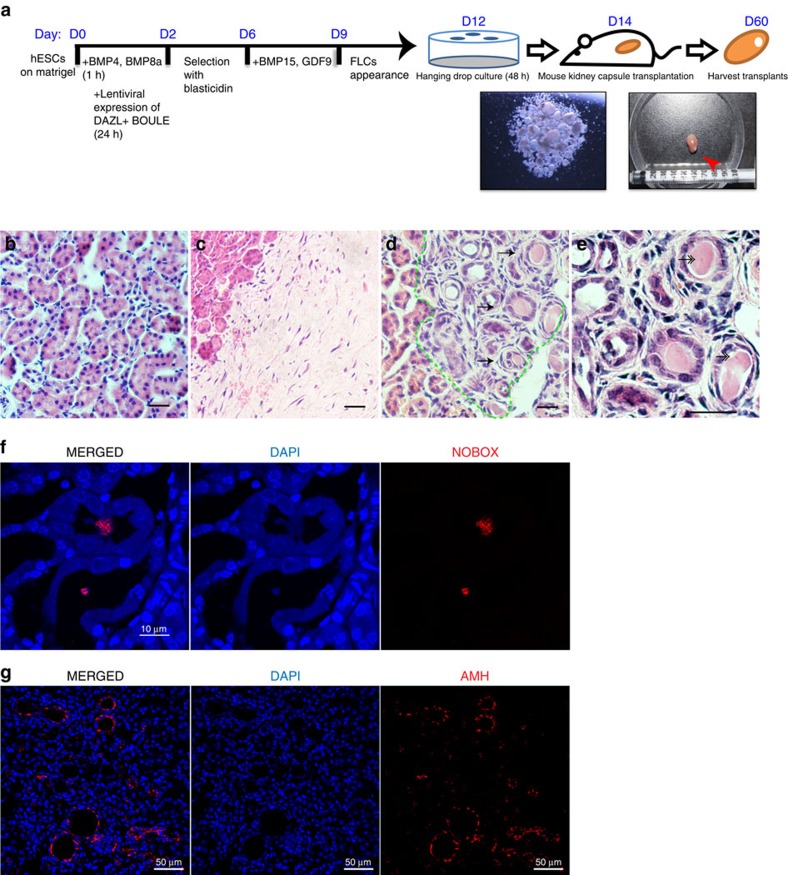
Figure 7. Existence of ovarian FLCs after transplantation into mouse kidney capsules.
(a) Schematic timelines and simplified protocols for the differentiation of the undifferentiated hESCs to the harvesting of the transplant from mouse kidney capsules. Red arrow: harvested transplant. Representative images of H&E staining of a kidney area next to the transplant (b), a control transplant containing differentiated hESCs without inducers (c), a transplant containing suspended FLCs as described in (d), and a magnified picture of FLCs in the transplant (e). Arrows: FLCs with primary follicle-like structures; double-headed-arrows: GV-like staining in the FLCs. (f) Immunofluorescent staining of NOBOX in the transplant showing colocalisation of NOBOX and nuclei in the middle of follicles. (g) Immunofluorescent staining of AMH in the transplant showing the AMH-positive cells localized to the surrounding cells of follicles. Bar, 50 μm in b–e,g; 10 μm in f. Three independent transplantation experiments were conducted.

