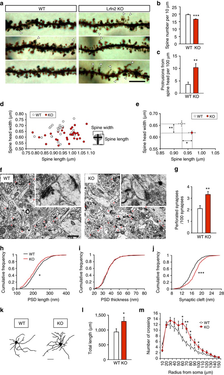
Figure 5. Morphological abnormalities of hippocampal synapses in Lrfn2 KO mice.
(a) Representative dendrite segments of Golgi-impregnated pyramidal neurons in the distal area of the hippocampal CA1 stratum radiatum from 8-week-old littermate WT and Lrfn2 KO mice. Scale bar, 5 μm. (b) Spine densities of WT and Lrfn2 KO neurons (WT, n=22 neurons from two animals, total 1,420 spines; KO, n=33 neurons from three animals, total 1,645 spines). Lrfn2 KO mice showed a slight reduction in the spine density (***P<0.001, t-test). (c) Number of spines having a protrusion from the spine head. (d) Mean width of the spine head is plotted against mean spine length for each neuron. Data from 22 WT neurons and 33 neurons in Lrfn2 KO. (e) Comparison of the mean of all spine widths plotted on the y-axis and the mean of all spine lengths on the x-axis (*P<0.05, **P<0.01, t-test). (f) Representative EM images of the hippocampal CA1 stratum radiatum. Red asterisks indicate synaptic contacts with a postsynaptic density, and insets show representative synapses at high magnification. Scale bar, 200 nm. (g–k) Quantitative analyses. The ratio of perforated synapses to total synapses (g) was larger in Lrfn2 KO mice (WT, n=11 sections from three animals; Lrfn2 KO, n=11 sections from three animals; *P<0.05, **P<0.01, t-test). (h–j) Cumulative frequency distribution of width of PSD length (h), PSD thickness (i) and synaptic cleft (j). Black lines indicate WT and red lines indicate Lrfn2 KO (WT, n=306 synapses; KO, n=301 synapses; *P<0.05, ***P<0.001, K-S test). Similar results were obtained in two independent experiments. (k–m) Morphological analysis of the cultured hippocampal neurons. (k) Representative tracings of MAP-2–immunostained neurons from WT and Lrfn2 KO mice at DIV10. (l) Quantification of the total dendrite length (WT, n=14 neurons; KO, n=12 neurons; *P<0.05, U-test). (m) The results of the Sholl analysis (WT, n=14 neurons; KO, n=12 neurons; *P<0.05, **P<0.01, t-test). Similar result was obtained in an independent experiment using DIV20 neurons (k–m).

