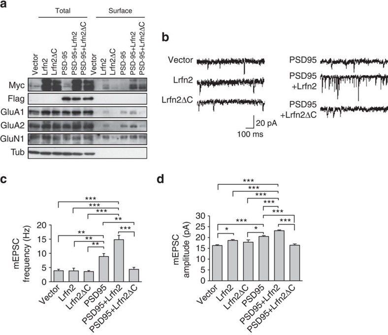
Figure 8. Interaction between Lrfn2 and PSD-95 facilitates the surface expression and synaptic strength of AMPAR.
(a) Biotinylation assay of cultured rat hippocampal neurons overexpressing Myc-Lrfn2, Myc-Lrfn2ΔC, Flag-PSD95, the combination of Myc-Lrfn2 and Flag-PSD95, or the combination of Myc-Lrfn2ΔC and Flag-PSD95. Neurons were transfected with the indicated vectors at DIV10 and biotinylated at DIV17. Immunoblots showed both total and biotinylated (surface) proteins of glutamate receptors. (b) Examples of AMPAR-mediated mEPSCs recorded from cultured rat hippocampal neurons. Neurons were transfected with the indicated expression vectors at DIV10 and analysed at DIV20–24. (c,d) (c) Summary of mEPSC frequency (n=5 neurons for each transfection except Lrfn2ΔC and PSD95 [n=6 neurons each]; *P<0.05, **P<0.01, ***P<0.001, ANOVA and post hoc Tukey–Kramer test). (d) Summary of mEPSC amplitude (Vector, n=286 events from five neurons; Lrfn2, n=268 events from five neurons; Lrfn2ΔC, n=159 events from six neurons; PSD95, n=457 events from five neurons; PSD95+Lrfn2, n=678 events from five neurons; PSD95+Lrfn2ΔC, n=202 events from five neurons; *P<0.05, **P<0.01, ***P<0.001, ANOVA and post hoc Tukey–Kramer test). The results are obtained using four independently cultured neurons.

