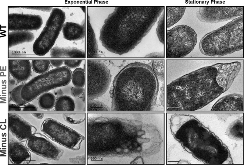
FIG 3.

Visualization of cellular ultrastructure by electron microscopy. Transmission electron microscopy of thin sections of WT and phospholipid-altered (minus-PE and minus-CL) cells grown in LB medium to exponential phase and stationary phase. Cells were grown aerobically in LB medium until they reached mid-logarithmic phase (OD600 of ∼0.3) or stationary phase (OD600 of ∼2). All specimens were fixed, embedded, ultrathin sectioned, and poststained before imaging on a JEOL 1400 electron microscope. Bars, 1 μm (left) and 200 nm (center and right).
