
Fig. 7.
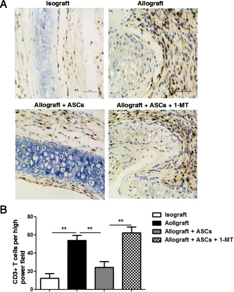
Effect of IDO inhibitor on CD3+ T cell infiltration in allografts with ASCs treatment. a Tissue sections from tracheal grafts on day 14 were immunostained with CD3 antibody and counterstained lightly with hematoxylin for viewing negatively stained cells. Cells stained with dark brown indicate CD3+ T-cell infiltration. b CD3+ T cells were quantitated from random fields under high magnification (400 ×). All data are expressed as mean ± SEM; n = 4 per group. **, p < 0.01