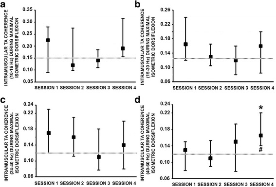
Fig. 5.

Intramuscular TA coherence estimated following incomplete SCI during the 4 repeated testing sessions. a. Median TA coherence analysed within the 10–16 Hz, b. 15–30 Hz, c. 24–40 Hz and d. 40–60 Hz frequency range during maximal isometric dorsiflexion from 20 subjects with SCI. Dotted line corresponds to the median non-injured group coherence value. *: p ≤ 0.05 with respect to session 1 and #: p ≤ 0.05 with respect to the non-injured control data. Further methodological information can be found in the analysis section of the methods. Data presented as median values with 25th and 75th percentiles
