
Fig. 1.
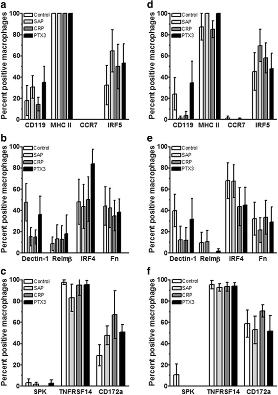
Expression of M1/M2 polarization markers on macrophages cultured with pentraxins. PBMC were cultured with either (a-c) 25 ng/ml M-CSF or (d-f) 25 ng/ml GM-CSF for 6 days and then SAP (10 μg/ml), CRP (10 μg/ml), or PTX3 (10 ng/ml) was added for an additional two days. PBMC were then air-dried, fixed, and stained by immunocytochemistry (ICC) with the indicated antibodies or irrelevant control antibodies. Following immunocytochemical staining, at least 100 macrophages were examined from at least 10 randomly selected fields, and the percentage of positive cells is expressed as the mean ± SEM (n = 4–5 separate donors)