
Fig. 2.
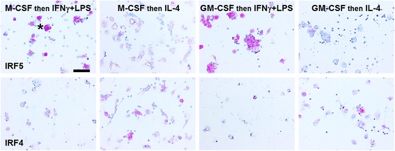
Expression of IRF5 (M1 marker) and IRF4 (M2 marker) on polarized macrophages. PBMC were cultured with 25 ng/ml of either M-CSF or GM-CSF for 6 days, and macrophages were then polarized for 2 days with either LPS + IFNγ or IL-4. Cells were then air-dried, fixed, and stained by ICC with antibodies. Positive cells are identified by red staining, and nuclei are counterstained blue. Bar is 100 μm. Asterisk indicates a cluster of macrophages stained with anti-IRF5 antibodies