Fig. 9.

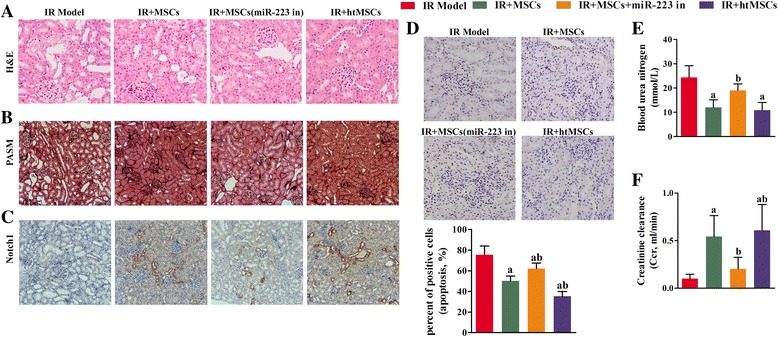
Perfusion of mesenchymal stem cells (MSCs) attenuated ischemia/reperfusion (IR)-induced damage to kidney tissue, and initiated Notch signaling in the mouse kidney. a Representative images of hematoxylin and eosin (H&E) stained tissue sections (×200). Administration of MSCs containing active miR-223 restored the integrity of cellular structures that had been damaged as a result of IR induction. b Representative images from periodic acid-silver methenamine (PASM) staining studies (×100 magnification). The collagen fibers are stained dark and the amount of kidney fibrosis was attenuated by perfusion with functional MSCs. c Representative images from immunochemical assays (×100 magnification); Notch1-positive cells are stained brown. Notch1 expression in the kidney cells of model mice was initiated by perfusion of MSCs transfected with a NC inhibitor or MSCs pretreated with hypoxia (htMSCs). d Representative images from TUNEL staining studies (×200 magnification). Apoptotic cells are stained brown, and their numbers were decreased after treatment with MSCs transfected with a negative control inhibitor or MSCs pretreated with hypoxia. Six fields were analyzed in the assay. e Blood urea nitrogen (BUN) assays showed that treatment with MSCs or htMSCs significantly decreased the levels of BUN compared to those in the IR group; however, a decrease in miR-223 expression reversed the reductions in BUN caused by MSCs or htMSCs. f The creatinine clearance rates were elevated in both the IR + MSC and IR + htMSC groups. The loss of miR-233 alleviated the increase in creatinine clearance caused by MSCs or htMSCs. a P < 0.05, versus the IR model group; b P < 0.05, versus the IR + MSC group. miR-233 in miR-233 inhibitor
