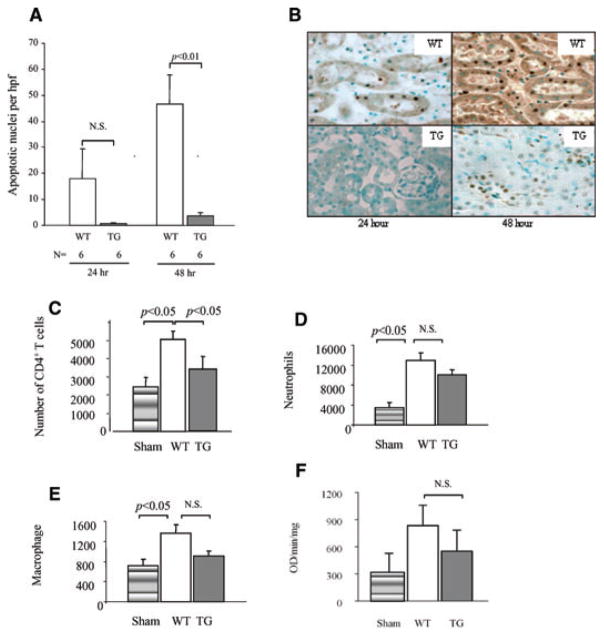
Figure 4. Apoptosis and CD4+ T-cell infiltrate are reduced in hCD39 kidneys after IRI.
(A) Sections of renal tissue were analyzed by TUNEL-stain and the number of apoptotic nuclei per high power field (hpf) were counted. Results are presented as mean ± SEM. N.S (not significant). (B) Representative sections of TUNEL-stained renal cortex at 24 and 48 h. Magnification 400×. (C–F) Mice were subjected to 30 min of ischemia and 3 h of reperfusion. Leukocytes were extracted from the kidneys and analyzed by flow cytometry. (C) CD4+ T cells. (D) Neutrophils. (E) Macrophages. (F) MPO activity assay was performed following 3 h of reperfusion. Data are representative of two independent experiments; n = 4 for each group in each experiment.

