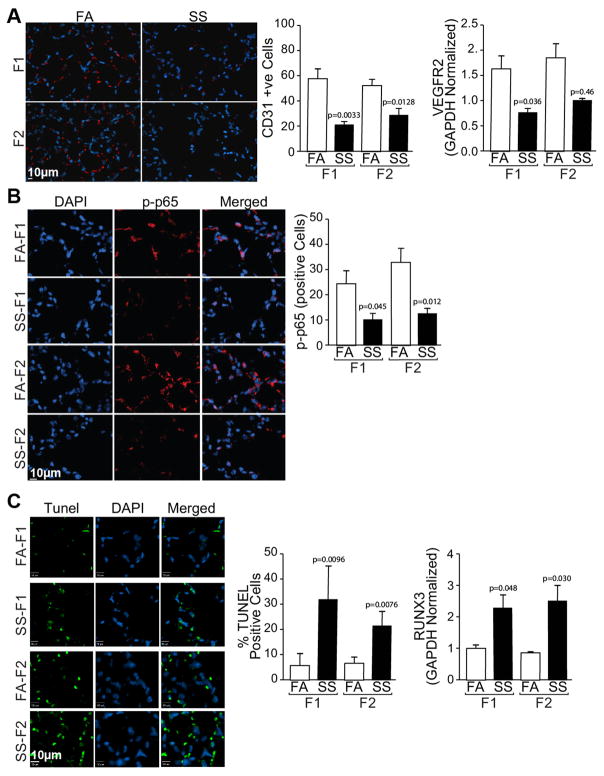
Fig. 4. Gestational SS-exposure downregulates angiogenesis, VGEFR2, p65-NFκB, and upregulates apoptosis in F1 and F2 lungs.
A: Lung sections were stained with anti-CD3 antibody (left anel) and the red florescent cells were quantitated microscopically (middle panel); expression of VEGEFR2 was done by qPCR analysis (right panel).B: Lung sections were stained with phosphorylated(p)-anti-p-65 antibody (left panel) and p-65-positive cells were quantitated (right panel). C: Lung sections were stained for TUNEL-positive cells using In Situ cell Death Detection kit (left panel) and quantitated microscopically (middle panel); RUNX3 levels were determined by quantitative RT-PCR using total lung RNA. Where indicated slides were counterstained with DAPI to visualize nuclei. Results are representative of animal responses from two different sets of inhalation exposures. Data are expressed as mean ± SD (n = 5–7/group. FA = filtered air, SS = secondhand cigarette smoke; F1: first generation; F2 second generation.

