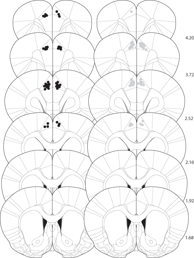
Figure 5.

Histological verification of cannula placements for rats used in experiments 1–4. Approximate locations of infusion tips, in the ACgx. The black dots (left panel) represent the location of the injector tips in experiments 1 and 2; the gray dots (right panel) the location of the injector tips in experiments 3 and 4. Placements are shown on coronal plates adapted from Paxinos and Watson (1998) with numbers indicating distance from bregma in millimeters.
