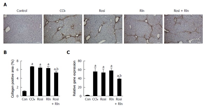
Figure 2.

Liver type I collagen content. A: Immunohistochemical staining of liver tissue from control (Con), fibrotic (CCl4), rosiglitazone (Rosi), serelaxin (Rln) or combination-treated (Rosi + Rln) mice. Bar: 100 μm; B: Type I collagen staining quantified; C: Type I collagen gene expression determined by qPCR. Data are expressed as mean ± SE, and analyzed by ANOVA (n = 5). aP < 0.05 vs Con; bP < 0.05 vs CCl4, Rosi, or Rln.
