
Figure 3. In vitro expression of specific meniscus ECM components in hMSC constructs.
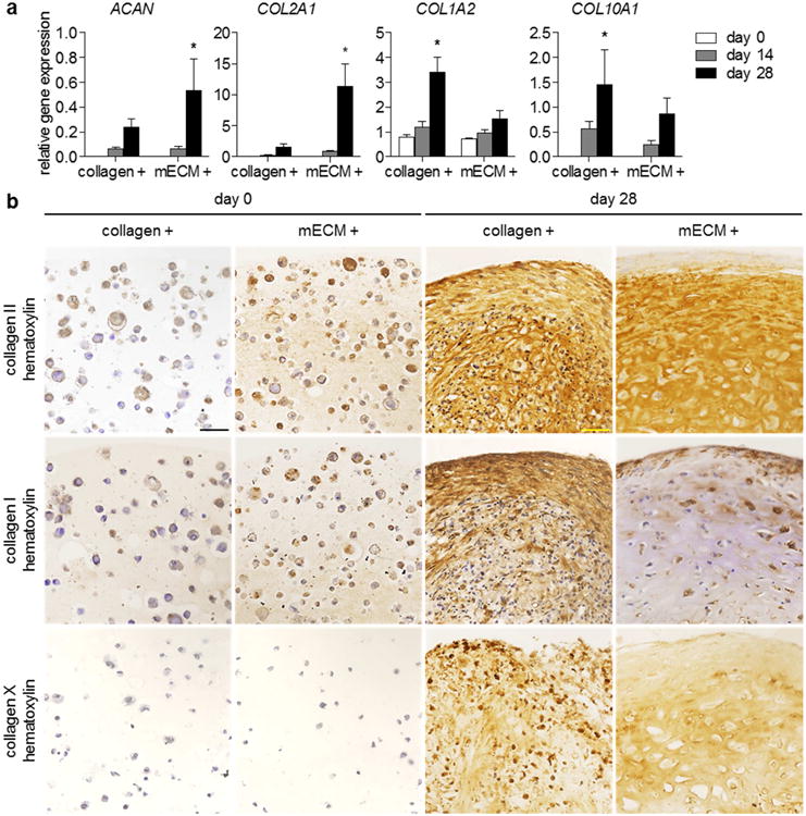
(a) Relative gene expression (2-ΔCT) of hMSCs encapsulated in type I collagen or mECM hydrogels, over 28 days of culture with TGF-β3 supplementation. Expression of aggrecan (ACAN) and type II collagen (COL2A1) were significantly greater at day 28 in mECM constructs, whereas type I collagen (COL1A2) and X (COL10A1) were significantly greater at day 28 in collagen constructs. ACAN: * p < 0.05 vs. mECM days 0, 14; n = 4. COL2A1: * p < 0.05 vs. mECM days 0, 14, collagen day 28; n = 4. COL1A2: * p < 0.05 vs. collagen days 0, 14, mECM day 28; n = 4. COL10A1: * p < 0.05 vs. collagen day 0; n = 4. (b) Immunohistochemistry of hMSC-mECM and collagen constructs. Intense staining for type II collagen appears at day 28 in both constructs, but only mECM hydrogel contained type II collagen initially. Positive staining for type I collagen is present in both collagen and mECM constructs at day 0, but staining is more prevalent at day 28 throughout collagen constructs. Collagen X does not appear in either hydrogel at day 0, but develops by day 28, although positive staining is present both intra- and extracellularly. Scale bar: 50 μm.