Figure 6.
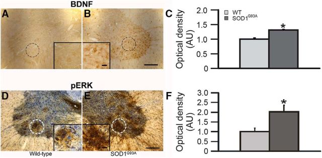
A–F, BDNF (A–C) and pERK (D–F) expression in putative phrenic motor neurons. Representative BDNF (A, B) and pERK (D, E) immunostaining in putative C4 phrenic motor neurons from wild-type (A, D) and SOD1G93A (B, E) rats; BDNF and pERK immunostaining are a brown reaction product. In D and E, neurons were counterstained with cresyl-violet (blue). The putative phrenic motor nucleus is circumscribed in A, B, D, and E, shown at higher magnification in insets. Densitometry was performed within these circumscribed areas. OD measurements demonstrated significant increases in BDNF (C) and pERK (F) expression in the putative phrenic motor nucleus of SOD1G93A (dark gray bar) rats compared with wild-type (WT; light gray bar) littermates. Values are means ± 1 SEM. *p < 0.05 versus wild-type rats. Scale bars: low magnification, 200 μm; high magnification, 50 μm.

