Figure 5.
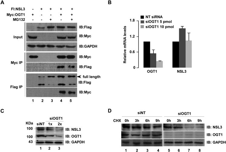
Stabilization of NSL3 by OGT1 was observed in cells. A, stabilization of NSL3 by OGT1 in 293T cells. Cells were co-transfected with FLAG-NSL3 and Myc-OGT1 as indicated with or without MG132 (10 μm; 12 h). 48 h later, co-IP was performed with prepared whole-cell lysates. Bound proteins were confirmed by Western blotting with anti-FLAG or anti-Myc antibody. B and C, expression level of NSL3 in siOGT1-treated 293T cells. mRNA and protein levels of NSL3 were measured using quantitative real time PCR (B) and Western blotting (C), respectively, in OGT1 knockdown 293T cells. D, expression level of NSL3 in OGT1 knockdown cells treated with CHX. OGT1 knockdown 293T cells were treated with CHX (3 μg/ml) for 0, 3, 6, and 9 h. Proteins in whole-cell lysates were detected using specific antibodies. IB, immunoblot; Fl, FLAG; NT, non-targeting.

