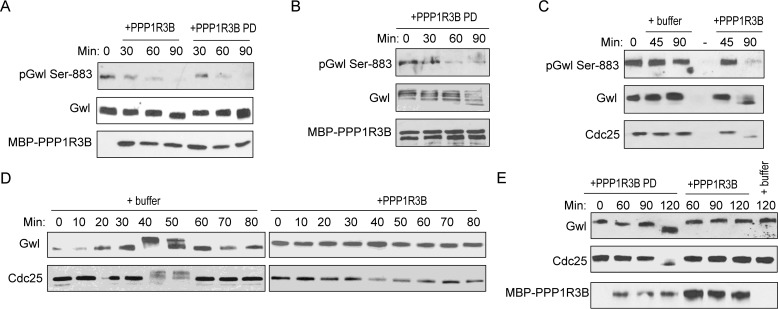
Figure 6.
PPP1R3B regulates mitotic entry and exit in Xenopus egg extracts. A, CSF extracts were diluted in a phosphatase buffer (50 mm HEPES, 100 mm NaCl, 2 mm DTT, 0.01% Brij 35, and 1 mm MnCl2) and incubated with MBP-PPP1R3B beads that were mock-treated or pulled down (PD) from interphase egg extracts. Extract samples at various time points were analyzed by immunoblotting for phospho-Gwl Ser-883, Gwl, and MBP. B, the MBP-PPP1R3B pulldown as in A was incubated with purified active Gwl for 30, 60, and 90 min. Dephosphorylation of Gwl Ser-883 was monitored by immunoblotting for phospho-Gwl Ser-883, Gwl, and MBP. C, CSF extracts were incubated with buffer or purified MBP-PPP1R3B. Extract samples at various time points were analyzed by immunoblotting for phospho-Gwl Ser-883, Gwl, and Cdc25C. D, cycling egg extracts were supplemented with buffer or purified MBP-PPP1R3B. Extract samples at various time points were analyzed by immunoblotting for Gwl and Cdc25C. E, CSF extracts were incubated with buffer (last lane) or MBP-PPP1R3B beads that were mock-treated or pulled down from interphase egg extracts. Extract samples at various time points were analyzed by immunoblotting for Gwl, Cdc25C, and MBP. The data in this figure are representative of three or more independent experiments.

