
Figure 3.
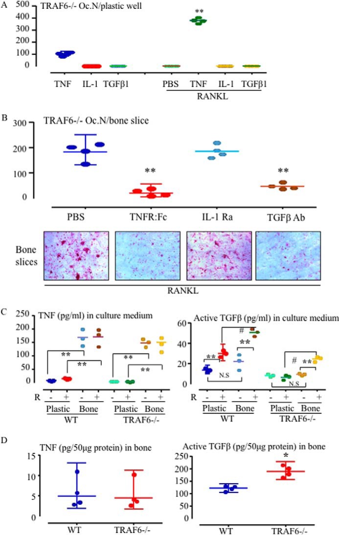
Either TNF or TGFβ1 mediates RANKL-induced OC formation from TRAF6−/− OCPs on bone. A, TRAF6−/− spleen cells were cultured with TNF, IL-1 (10 ng/ml), or TGFβ1 (2 ng/ml) or with each of them in combination with RANKL plus M-CSF in 96-well plastic plates for 5 days to generate OCs. TRAP+ OCs were counted after TRAP staining. **, p < 0.01 versus cells treated with TNF alone. Oc.N, OC number. B, spleen cells from 11-day-old WT and TRAF6−/− mice were seeded on bone slices in 96-well plates and cultured with RANKL and M-CSF plus the TNF inhibitor TNFR:Fc, an IL-I receptor antagonist (IL-1Ra), or a TGFβ-neutralizing Ab for 5 days. TRAP+ OCs were counted on bone slices after TRAP staining. **, p < 0.01 versus PBS-treated culture. C, 2 × 10⁵ spleen cells from 11-day-old WT and TRAF6−/− mice were seeded in 96-well plates on plastic or on bone slices and treated with PBS (P) or RANKL (R) plus M-CSF for 5 days. Levels of TNF (left panel) and the active form of TGFβ (right panel) in the culture media were tested by ELISA. **, p < 0.01 versus the respective level in medium from cells cultured on plastic (n = 3/group); #, p < 0.05; N.S, no significant difference between the two groups. D, the lower extremities of 10-day-old TRAF6−/− and WT mice were homogenized in 1 ml of tissue protein extraction reagent containing a proteasome inhibitor mixture. 50 μg of tissue lysate protein from each sample was used to test the levels of TNF (left panel) and the active form of TGFβ1 (right panel) by ELISA (n = 4). All experiments were repeated at least twice with similar results.