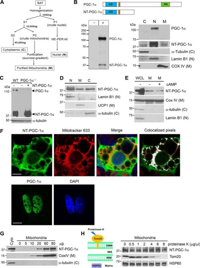
Figure 1.

NT-PGC-1α is present in brown adipocyte mitochondria. A, schematic of subcellular fractionation. Brown adipose tissues were isolated from C57BL/6J mice exposed to 4 °C for 5 h and subjected to subcellular fractionation. B, presence of NT-PGC-1α in mitochondria. Top panel, schematic of the PGC-1α and NT-PGC-1α proteins. AD, transcription activation domain; LxxLL, nuclear receptor interaction motif; RS, arginine/serine-rich domain. Bottom left panel, PGC-1α-HA and NT-PGC-1α-HA were expressed in COS-1 cells and immunoblotted with a highly specific anti-PGC-1α monoclonal antibody (9). Bottom right panel, endogenous PGC-1α and NT-PGC-1α in BAT isolated from cold-exposed mice were immunoblotted with the same anti-PGC-1α antibody. The cytosolic (C), nuclear (N), and mitochondrial (M) markers were detected in their respective fractions. C, expression of NT-PGC-1α in PGC-1α−/− brown adipocytes. WT brown adipocytes expressing an empty vector and PGC-1α−/− brown adipocytes stably expressing NT-PGC-1α or an empty vector by retrovirus-mediated gene transfer were treated with 0.5 mm dibutyryl cAMP for 4 h. D, presence of NT-PGC-1α in brown adipocyte mitochondria. PGC-1α−/− brown adipocytes stably expressing NT-PGC-1α were treated with dibutyryl cAMP and subjected to subcellular fractionation. E, cAMP-induced signaling does not regulate mitochondrial targeting of NT-PGC-1α. PGC-1α−/− brown adipocytes stably expressing NT-PGC-1α were treated with vehicle or dibutyryl cAMP for 4 h. WGL, whole cell lysates. F, localization of NT-PGC-1α and PGC-1α in PGC-1α−/− brown adipocytes. NT-PGC-1α-HA and PGC-1α-HA were expressed in PGC-1α−/− brown adipocytes by adenovirus-mediated gene transfer and analyzed for cellular localization by indirect immunofluorescence with anti-HA antibody. Colocalized pixels were analyzed using an ImageJ analysis tool and are displayed in white on an RGB overlay image. Scale bars = 23 μm. G, correlation of the amounts of mitochondria and the protein levels of NT-PGC-1α. Mitochondrial enrichment of NT-PGC-1α was analyzed in increasing amounts of purified brown adipocyte mitochondria. H, mitochondrial NT-PGC-1α is protected from proteinase K digestion. Purified mitochondria (60 μg) were treated with increasing amounts of proteinase K.
