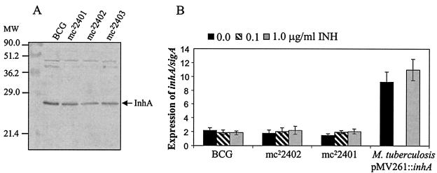
FIG. 3.
InhA expression in ndh mutants. (A) Western blot analysis. Detection of InhA in total protein extracts obtained from the different M. bovis BCG strains using rabbit anti-InhA antibodies raised against the M. tuberculosis InhA protein. (B) inhA mRNA levels in M. bovis BCG strains and the M. tuberculosis pMV261::inhA strain without addition of INH and after 4 h of incubation in the presence of 0.1 or 1.0 μg of INH/ml. inhA values were normalized using sigA levels as reference (26). Each value is the average of three culture replicates, each of which was evaluated three times. Error bars correspond to 95% confidence intervals. As a positive control of inhA overexpression, the M. tuberculosis strain harboring pMV261::inhA is shown (26), tested only without antibiotic and 1.0 μg of INH/ml.

