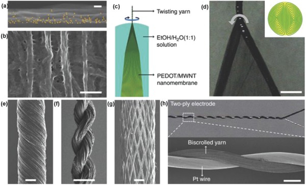
Figure 25.

a) An EDAX (energy‐dispersive analysis by X‐ray) map of the cross‐section of a PEDOT‐infiltrated two‐layer CNT sheet stack. Scale bar, 50 nm. b) SEM image of the side of a PEDOT‐coated MWNTs nanomembrane. Scale bar, 200 nm. c) Schematic illustration showing the fabrication of a biscrolled PEDOT/MWNT yarn. d) Optical microscope image of the spinning wedge, which shows the wedge edges being twisted to form a dual‐Archimedean scroll yarn, which is schematically illustrated in the inset. Scale bar, 200 mm. e) SEM image of a biscrolled yarn with bias angle. Scale bar, 10 mm. f) SEM image of two PEDOT/MWNT yarns plied together. Scale bar, 50 mm. g) SEM image of a braided structure containing 32 biscrolled yarns. Scale bar, 100 mm. h) Two SEM images of a PEDOT/MWNT biscrolled yarn that is plied with a 25 mm Pt wire. Scale bar, 40 mm. Reproduced with permission.506 Copyright 2013, Nature Publishing Group.
