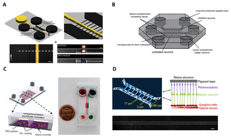
Figure 4.
Applications of neuron and ocular synapses studies A) Schematic of the microfluidic chip used for the visualizations and manipulation of synapses, where flow direction is achieved with the use of negative pressure thus by preventing diffusion into the microgrooves. The use of Alexa Fluor 488 allows for the observation of the difference in profile between perfusion and the washout step108. B) Experimental setup of the 3 compartment microfluidic device used for synaptic competition. The use of microgrooves on the side chambers and ‘target’ neurons in the centre establishes the synaptic competition between axons to reach the central chamber111. C) Schematics and actual image of neuron-astrocytes interaction microfluidic chip using food dyes to show the chambers and channels. This platform used for the first time simultaneously a combination of PEG, (polyethylene glycol), compartmentalized chambers and gen etically encoded calcium indicators for the analyses of neuron-neuron and neuron-astrocytes interaction88. D) Microfluidic device schematics for retinal synapse regeneration above the retinal structure mimicked with the use of two chambers connected by 108 microchannels. Scale bar, 200μm116. 4A. Reproduced from Ref 108 with permission from Elsevier. 4B. Reproduced from Ref 111 with permission from Elsevier. 4C. Reproduced from Ref 88 with permission from Nature Publishing Group. 4D. Reproduced from Ref 116 under a CC-BY license through Nature Publishing Group.

