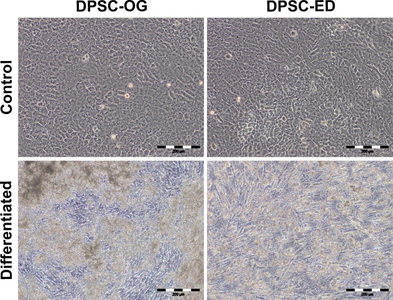
Figure 6. Morphology of DPSC-OG and DPSC-ED at day 21 of osteoblastic differentiation.
DPSC was cultured in proliferation medium (control) and osteoblastic differentiation medium (differentiated). After 21 days, there was a decrease in the confluency of DPSC-ED in differentiation medium as compared to control. No visible change in the confluency was detected between DPSC-OG in the control and differentiation medium. Scale: 200 µm.

