
Figure 3. JUN is activated by MEK signaling in response to ER stress.
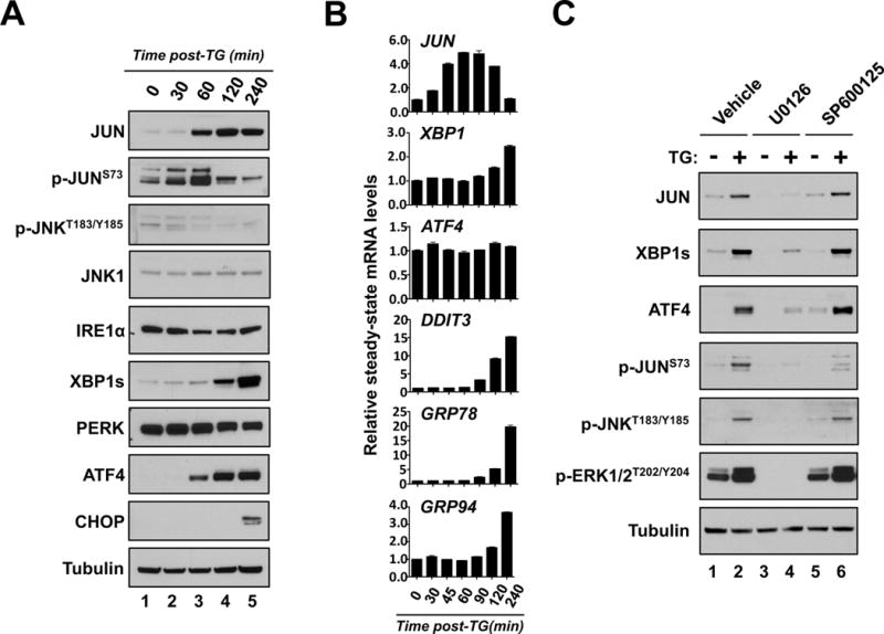
A & B. THP-1 cells were treated with TG and subsequently harvested for protein expression (A.) or (B.) mRNA analysis at the indicated times. A. Protein lysates from each of the indicated time points were subjected to western blot analysis using the indicated antibodies. B. Real-time qPCR analysis of JUN, XBP1, ATF4, CHOP, GRP78 and GRP94 expression at the indicated times. C. THP-1 cells were treated without (−) and with TG (+) in combination with vehicle, 10uM U0126 or 10uM SP600125 for 3 hours. Lysates from each condition were then analyzed by western-blot with indicated antibodies.