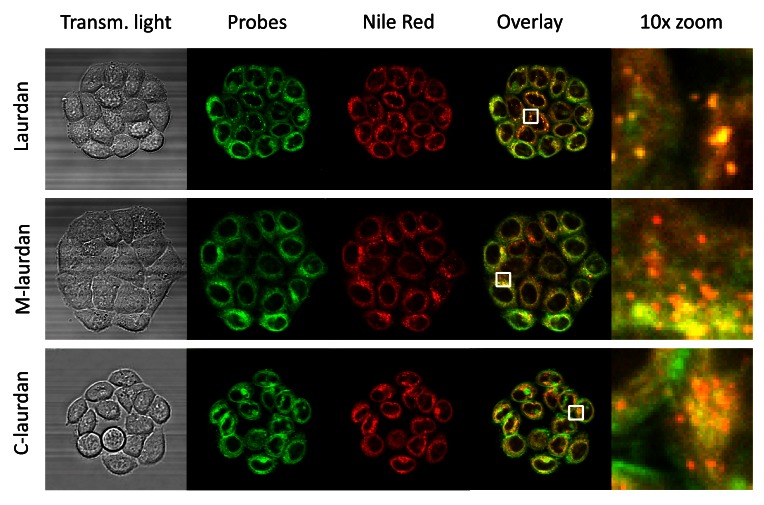
Figure 3. The intra-cellular vesicles labelled by Laurdan co-localize perfectly with Nile Red labelling, which labels lipid droplets.
Hela cells were grown on glass coverslips for four days before double-labelling with Nile red and either Laurdan (first line), M-laurdan (second line) or C-laurdan (third line), as described in M&M. The live cells, kept at 37°C, were then imaged over 30 channels: 29 fluorescence channels spanning from 420 to 700 nm, plus transmitted light (First column). The stacks were then used to perform spectral deconvolution with the poisson NMF pluggin, using the spectra acquired with cells labelled with only one probe as references. The second column: shows the signals allocated to the Laurdan-family probes, artificially coloured in green. The third column shows the signals allocated to Nile red, artificially coloured in red. The fourth column shows an overlay of the two signals, and the fifth column shows 10× zooms corresponding to the white squares in the fourth column. Width of the squares: 10 µm.

