Fig. 2.
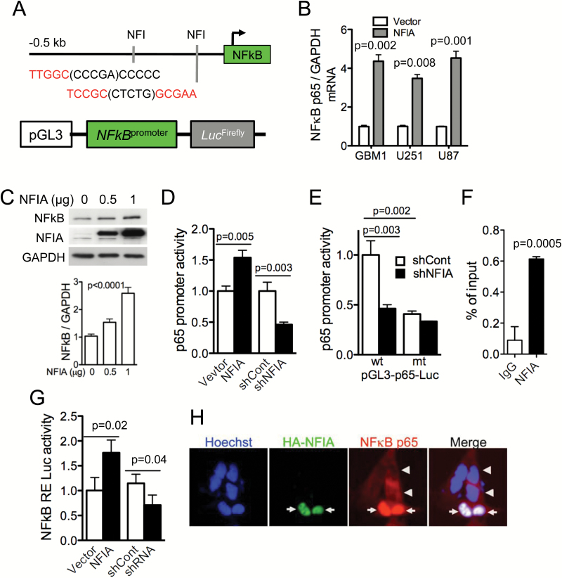
NFIA increases NFκB p65 transcription and induces NFκB activity and nuclear translocation. (A) Putative NFIA binding sites in the human NFκB p65 promoter. (B) Relative mRNA expression of NFκB p65 to GAPDH in GBM cells expressing NFIA or vector was determined by RT-PCR and densitometric analysis. (C) Immunoblots of whole cell lysates from GBM1 cells transiently transfected with the increasing amount of a lentivector encoding NFIA (bottom; densitometry). (D) Luciferase activities were measured 48 h after transfection of NFκB p65 luciferase reporter into U87 GBM cells expressing NFIA, shNFIA, or controls (vector or shCont); n = 3. (E) Relative luciferase activity of pGL3 wild-type (wt) or mutant (mt) NFκB p65 promoter transfected into U87 GBM cells expressing shNFIA or shCont similar to the left panel. (F) ChIP-qPCR assays on GBM cells using anti-NFIA or control immunoglobulin G. (G) Luciferase assay with the 3x κB-luc-reporter in U87 cells transfected with NFIA constructs (NFIA, vector only, shNFIA, or shCont). (H) NFIA promotes nuclear translocation of NFκB p65. Immunofluorescence staining of GBM1 cells expressing HA-NFIA or vector control with Hoechst (nuclei; blue), anti-HA (green), anti-p65 NFκB (red): arrows: nuclear p65 in HA-positive cells, arrowheads: cytoplasmic p65 in HA-negative control cells.

