Figure 1.

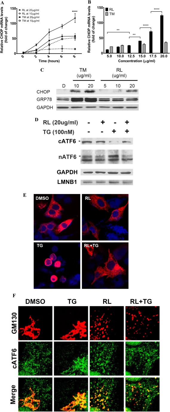
Effects of anti‐HIV drugs on ER stress response and cellular localization of ATF6. (A) Time course of mRNA of ER stress marker CHOP. HepG2 cells were treated with RL or TM. (B) Dose response of CHOP mRNA in the RL‐ or TM‐treated cells. (C) GRP78 and CHOP protein expression in TM‐ and RL‐treated cells. HepG2 cells were treated with TM or RL for 8 hours at different concentrations; D, DMSO as vehicle control. (D) Effect of RL on the processing of ATF6 protein in HepG2 cells. Cells were transfected with FLAG‐ATF6 for 24 hours and treated with either DMSO or RL at 20 μg/mL for 2 hours followed by treatment with 100 nM TG for 2 hours. GAPDH and LMNB1 were used as loading controls for cytoplasmic and nuclear proteins, respectively. (E) Blockage of TG‐induced nuclear ATF6 by RL. Confocal images (×630) showing nuclear localization of ATF6 in the cells expressing ATF6. (F) RL‐induced dispersal of ATF6 and Golgi components. Confocal images (×1000) showing changes of colocalization and dispersal of ATF6 (green) and GM130 (red) in the cells exposed to the drugs. **P < 0.01; ****P < 0.0001 compared to control or indicated otherwise; n = 5‐7. Error bars indicate standard error mean (SEM). Abbreviations: cATF6, cytoplasmic ATF6 precursor; DMSO, dimethyl sulfoxide; GAPDH, glyceraldehyde 3‐phosphate dehydrogenase; LMNB1, lamina B1; nATF6, activated nuclear ATF6.
